
Imaging flow cytometers combine the resolution of fluorescence microsopy with the throughput of flow cytometry, allowing researchers to analyze physical features of tens of thousands of cells or EVs in a few minutes. However, building an imaging flow cytometer requires more complex optics and higher performance fluidics than traditional flow cytometers. As a mechanical engineering on the R&D team, I helped to develop the next generation of this technology for Luminex.

Luminex imaging flow cytometers work by hydrodynamically focusing cell samples into a single-file line and take high-resolution fluorescent images at rates of 1000+ cells/second
Design
Mathematical Modeling / CAD / Architecture and System Design
I was responsible for the development of the fluidic system. I developed mathematical models and used them to develop system architectures to hit performance targets. I worked with suppliers to find pumps, valves, and sensors for those architectures and design components in SolidWorks to connect them together and manage flow control.
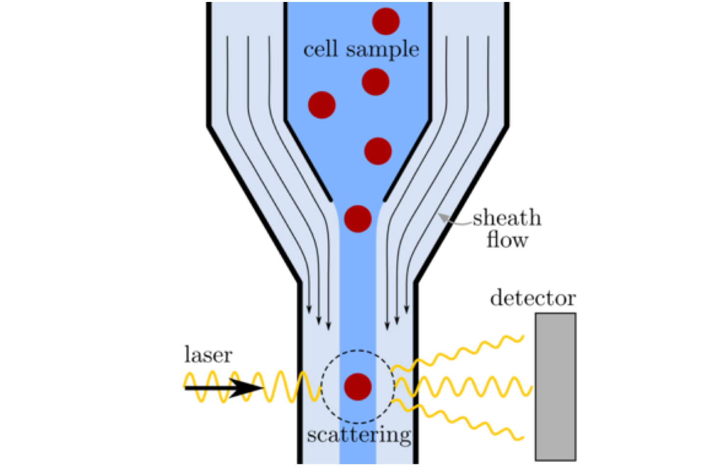
Drawing of the heart of flow cytometer: the flow cell. Tightly controlled sheath flow focuses the sample flow to permit individual cells to be imaged.
Testing
Test Design / Test Systems / Analysis
As part of my role I ran characterization and analysis of system fluidics, and construct test systems running LabView to run tests I developed. I analyzed the resulting data using Python in Jupyter notebooks.
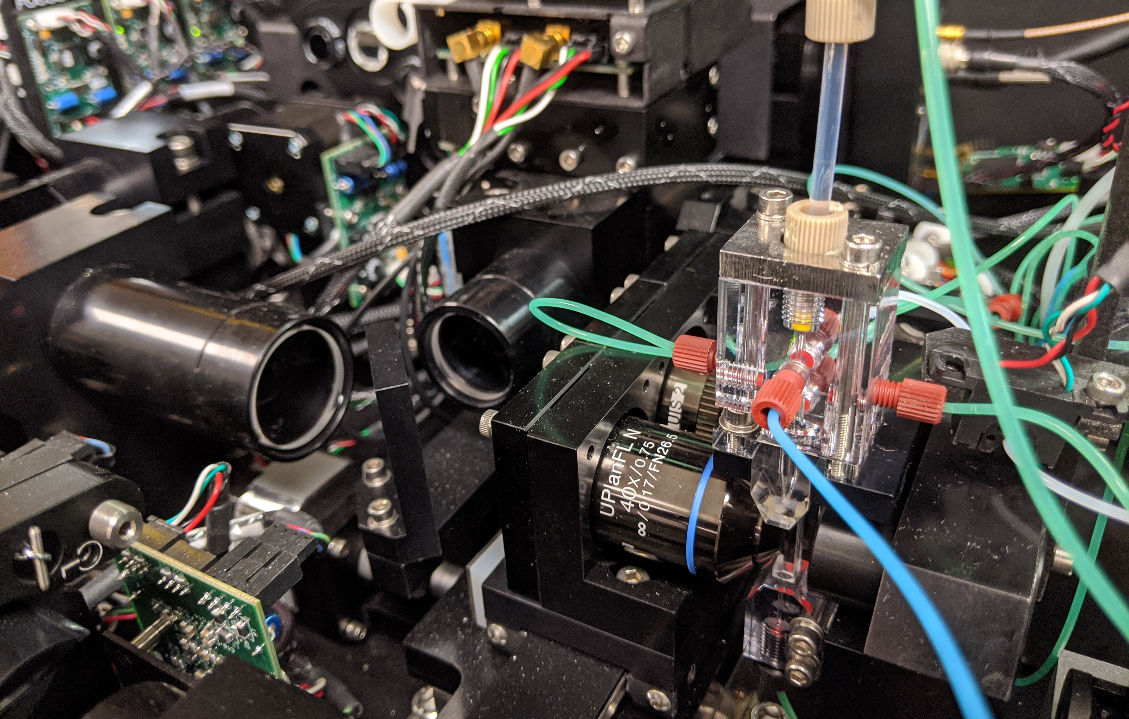
Flow cell and condenser of the Luminex ImageStream X Mk2
Manufacturing
Design Transfer / Manufacturing Verification
I have developed drawings and written manufacturing instructions for dozens of components and assemblies to enable transfer to manufacturing, and built a pump characterization tool to verify performance on the manufacturing line.